
Opening or hole in bone that permits the passage of nerves and blood vessels. The region between mental foramens is considered as a zone of choice for implants.

Visibility Of Mandibular Anatomical Landmarks In Panoramic Radiography A Retrospective Study Semantic Scholar
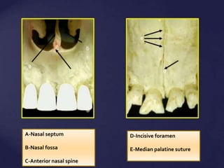
The incisive foramen is an opening in the midline of the palate just posterior to the central incisors.

. This program is Normal Radiographic Anatomy of Maxillary Periapical Projections This unit presents an introductory identification of the normal anatomy seen in maxillary periapical radiographs. The incisive nerve innervates the anterior palatal soft tissues. Although occasionally observed in radiographic examinations of the incisor area of the maxilla nasopalatine duct cysts were.
3B can be seen leading to the incisive fora-. A foramen is a n. Its precise location is.
Incisive foramen is the opening of the incisive canal located immediately behind the maxillary central incisors. Several authors have reported different dimensions of radiolucency as diagnostic of. Singer DDS 2123055674 srs2columbiaedu.
This chapter presents the major landmarks commonly found on conventional dental x-ray images. Question was removed from public access Tic douloureux is synonymous with If hypothyroidism occurs in the adult it can be associated with The radiographic image of the incisive foramen is located between the roots of the maxillary Recurrent herpes labialis is Presence of periodontal pockets increased tooth mobility pus formation and bad. Broad shallow scooped-out or depressed area of bone.
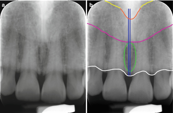
On periapical x-ray images the incisive foramen is located in the midline between the roots of the central incisors. Our goal is to evaluate identification of MIC by both panoramic radiograph PAN and cone-beam computed tomography CBCT. The incisive foramen is important because it is a potential site of cyst formation.
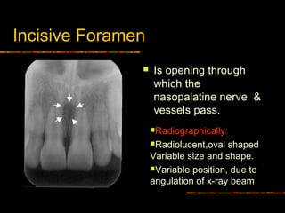
Here a posterior-occlusal view of a skull demonstrates the incisive foramen. In the human mouth the incisive foramen also known as. Opening or hole in bone that permits the passage of nerves and blood vessels.
Hollow space cavity or recess in bone. NASOPALATINE duct cysts are cysts which form in the incisor canal region of the maxilla and originate in the nasopalatine duct or its remnants. The mental foramen MF is located in the anterolateral region of the mandible body and through it passes the mental nerve and vessels.
Anterior palatine foramen or nasopalatine foramen is the opening of the incisive canals on the hard palate immediately behind the incisor teethIt gives passage to blood vessels and nerves. On a _____ periapical radiograph the incisive foramen appears as a small ovoid or round _____ area located between the roots of the central incisors. Mean canal length was 1863 235 mm and males have significantly longer incisive canal than females.
These cysts have no direct relationship to the teeth but in their growth may encroach upon the incisor apices. The incisive foramen is situated within the incisive fossa of the maxilla. All of the above F.
E incisive foramen f median palatal suture b a d c facial view palatal view e f Landmarks in the Maxilla Incisive foramen Median palatine suture Pterygoid plates Pterodactyl gr. PDF On Jan 1 2020 K. As age of the subjects increased incisive foramen diameter and incisive canal length were.
The mean width of bone anterior to the incisive canal was 632 143 mm. None of the above. However complications may arise due to an extension anterior to the mental foramen that forms the mandible incisive canal MIC.
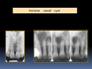
The presence of the cyst is presumed if the width of the foramen exceeds 1 cm or if enlargement can be demon-strated on successive radiographs. The nasopalatine duct cyst occurs in the nasopalatine or incisive canal and it may be difficult to decide on a radiograph whether radiolucency in that area is a cyst or a large incisive foramen. Sharp thornlike projection of bone.
In some radiographsB the incisive canal Fig. A working knowledge of normal anatomy of the oral-facial region as it appears on radiographs is essential in assessing accurately the information. Veena and others published Appreciation of Incisive Foramen in Intraoral Periapical Radiographs - A Comparative Radiographic Study.
The incisive foramen provides the exit of the nasopalatine nerve and artery from the palatine bone. Intraoral Radiographic Anatomy Steven R. The incisive foramen is used as an.
Median palatal suture D.

6 Essentials Of Dental Radiographic Analysis And Interpretation Pocket Dentistry

Maxillary Anterior Landmarks Intraoral Radiographic Anatomy Continuing Education Course Dentalcare Com

Periapical Radiograph 1 Year After Treatment Bone And Teeth Showing Download Scientific Diagram
Mouth Incisive Canal Cyst Professional Radiology Outcomes

Visibility Of Mandibular Anatomical Landmarks In Panoramic Radiography A Retrospective Study Semantic Scholar

The Appearance Of The Mental Foramen On Panoramic Radiographs Download Scientific Diagram
Normal Radiographic Anatomical Landmarks

Pdf The Evaluation Of Visibility Of Mandibular Anatomic Landmarks Using Panoramic Radiography Semantic Scholar

Measurement Of Nasopalatine Canal Length A Incisive Foramen Diameter Download Scientific Diagram
Radiographic Anatomical Landmarks By Dr Armaan Singh

Opg Showing Incisive Foramen And Mental Foramen Download Scientific Diagram
Normal Anatomical Landmarks In Dental X Rays And Cbct Springerlink

Panoramic Radiograph Showing Extension Of The Mental Nerve Beyond The Download Scientific Diagram

Figure 2 Assessment Of The Mandibular Incisive Canal By Panoramic Radiograph And Cone Beam Computed Tomography

Normal Radiographic Anatomical Landmarks

6 Essentials Of Dental Radiographic Analysis And Interpretation Pocket Dentistry

An Example Of A Large Incisive Canal Mesial To The Mental Foramen The Download Scientific Diagram
Intra Oral Radiographic Anatomical Landmarks
Normal Radiographic Anatomical Landmarks